
Pressure, Wounds, and Drains: Where This Book's Harm Lives
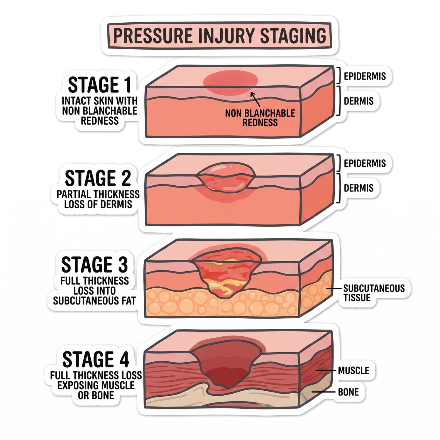
The stages, in current terminology
- Stage 1 — Intact skin with localized non-blanchable erythema. In darkly pigmented skin the change may present as a difference in color, temperature, firmness, or sensation rather than as redness.
- Stage 2 — Partial-thickness loss of skin with exposed dermis. The wound bed is viable, pink or red, and moist, sometimes presenting as an intact or ruptured serum-filled blister. No slough, eschar, or granulation tissue.
- Stage 3 — Full-thickness skin loss with adipose visible in the ulcer. Granulation tissue and rolled edges are often present, and undermining or tunneling may occur. Fascia, muscle, tendon, ligament, cartilage, and bone are not exposed.
- Stage 4 — Full-thickness skin and tissue loss with fascia, muscle, tendon, ligament, cartilage, or bone exposed or directly palpable.
- Unstageable — Full-thickness loss whose depth is obscured by slough or eschar. It will prove to be a Stage 3 or a Stage 4 once the base is visible.
- Deep tissue pressure injury — Persistent non-blanchable deep red, maroon, or purple discoloration of intact or non-intact skin.
Staging terminology is revised periodically. Verify every definition and every term on this page against the current National Pressure Injury Advisory Panel staging system before use.
The three staging rules
Pressure injuries are never reverse staged as they heal, so a healing Stage 4 is a healing Stage 4. A medical device related pressure injury is staged with this same system. A mucosal membrane pressure injury cannot be staged at all. Stable, dry, adherent eschar on an ischemic limb or heel is not softened or removed.
Wound assessment language
Describe rather than diagnose. Record location, measured length, width, and depth. Locate undermining and tunneling by clock position, with twelve o'clock toward the patient's head. Record wound bed tissue, exudate amount and character, wound edges, periwound skin, odor, and the patient's pain.
Dressings, matched to the wound
Dry gauze for packing and mechanical debridement. Transparent film for a dry, shallow, low-exudate wound. Hydrocolloid to hold moisture on a clean granulating wound. Hydrogel to donate moisture to a dry bed. Foam and alginate to absorb heavy exudate, with alginate reserved for the wettest wounds. The rule is simple: a wet wound needs absorption, a dry wound needs moisture.
Drains
A Penrose is open and passive, and the saturated dressing is the record of its output. A Jackson-Pratt bulb and a Hemovac evacuator are closed systems: empty, measure, and recompress each time, because a drain left uncompressed has stopped suctioning.
The stop-and-call list
- Dehiscence or evisceration. Cover with sterile saline-moistened gauze, position low Fowler's with the knees bent, keep the patient nothing by mouth, and call the surgeon. Never attempt to reinsert tissue.
- A stoma turning dusky, purple, or black.
- A sudden stop in drain output with rising pain and distention.
- A sudden change to bright red bleeding in a drain.
- New non-blanchable discoloration under any device.
Memory hook1 INTACT · 2 DERMIS · 3 FAT · 4 FLOOR.
Margin notedescribe it, don't diagnose it.
Watch outan unstageable wound is never assigned a number by guessing at its depth. It stays unstageable until the base is seen.
TakeawayStage by what tissue is visible, never by how bad it looks, and never backward.
Nutrition values in healing → BOOK 07 · P9. Vascular and diabetic contributions → BOOKS 10, 11.