
The coronary tree decides which wall dies
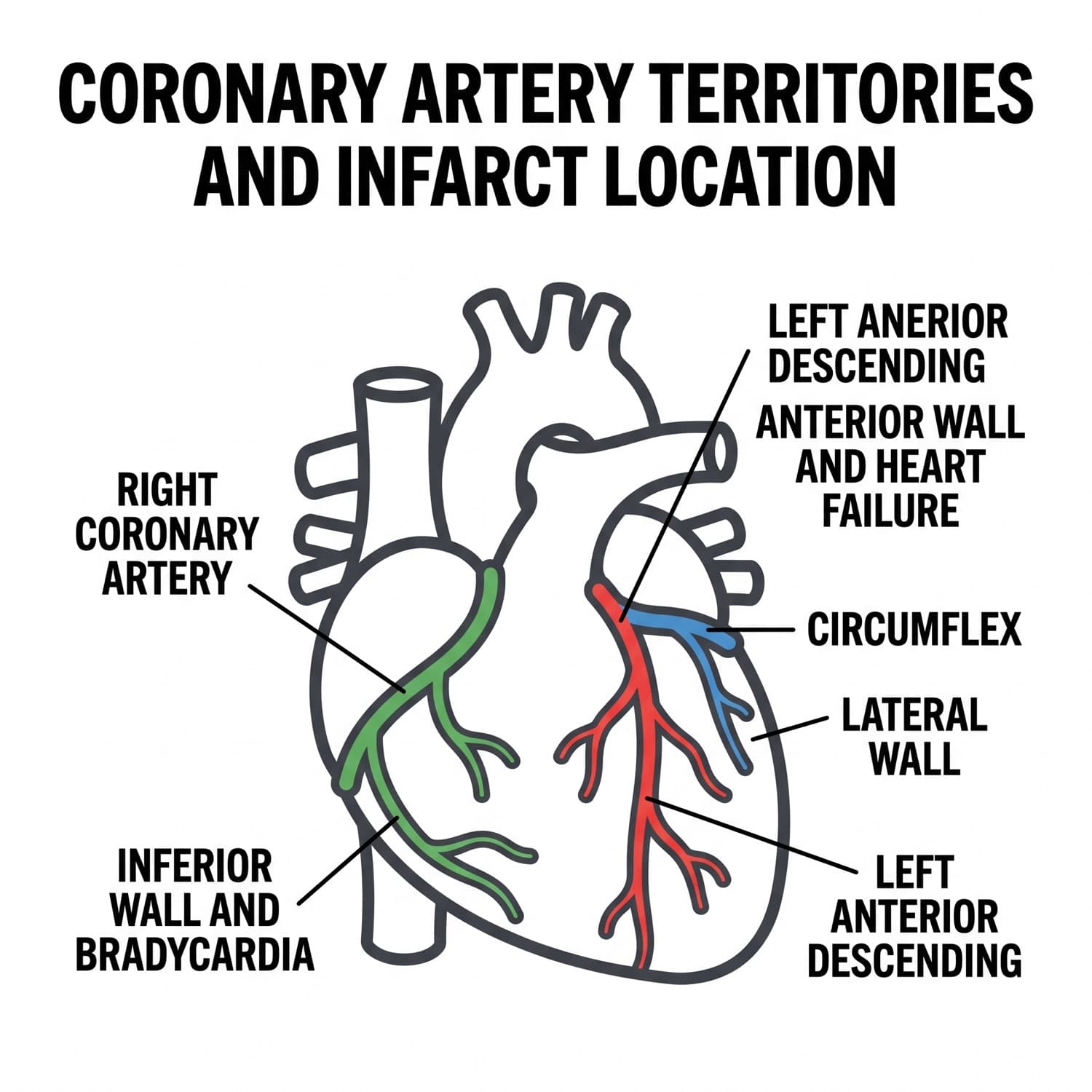
The pump has its own supply line. Which branch closes predicts which wall dies, and the wall predicts which complication walks in the door. Read the territory off the report, then assess for what that territory is known to do.
Panel 1 · Right coronary artery
- Feeds the inferior wall and, in most people, the sinoatrial and atrioventricular nodes.
- Expect bradycardia, first-degree block, Mobitz type I, and complete block.
- Expect right ventricular involvement, which makes the patient dependent on filling pressure.
- Leads II, III, and aVF carry the change, with reciprocal depression in I and aVL. A right-sided tracing is obtained when the inferior pattern appears.
Panel 2 · Left anterior descending artery
- Feeds the anterior wall and the interventricular septum, so it supplies the largest share of working muscle.
- Expect the biggest loss of contractility: new crackles, a third heart sound, falling output, and ventricular ectopy.
- Leads V1 through V4 carry the change.
- If perfusion collapses despite the pump support ordered, that is a shock state and it is escalated at → BOOK 09 · P9.
Panel 3 · Circumflex artery
- Feeds the lateral wall, with changes in I, aVL, V5, and V6.
- Often quieter on the monitor and easier to miss, which is exactly why the exam uses it to test serial assessment rather than pattern recognition.
- Also supplies the atrioventricular node in a minority of people, so nodal findings do not rule it out.
The days two to seven inset. Mechanical complications cluster after the acute hours, when necrotic tissue is at its softest. Papillary muscle rupture announces itself as a new loud murmur with sudden pulmonary congestion. Septal rupture adds a new murmur with an abrupt fall in forward output. Free wall rupture presents as tamponade physiology. All three are reported immediately.
Memory hookINFERIOR MEANS FLUIDS, NOT NITRATES. Registered as B8-INF-FLUID.
Tick box: can I name the artery from the wall, and the wall from the artery?
No infarct definitions here; the spectrum is at → BOOK 08 · P5. No marker values; → BOOK 07 · P4.
Takeawaythe artery predicts the complication, so the wall on the report tells you what to assess next.